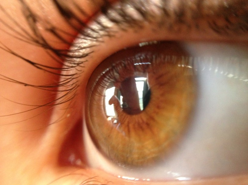
We often focus on the brain when it comes to memory impairment, but a new study has found that looking at retinas’ patterns of scattering light may give clues to diagnosing Alzheimer’s disease early on.
The retina is a layer of tissue at the back of the eye that receives light and then sends it on to the brain to make sense of the incoming visual messages. Because the retina is connected to the brain through the optic nerve and since proteins called “beta-amyloids” are often an early indicator of Alzheimer’s disease when found in the brain, scientists turned to the retina to search for clues to early diagnosis.
A team of researchers from the University of Minnesota hypothesized that, if small clumps of these proteins were present in a subject’s retina, light would scatter in a unique way. This could indicate the beginning of Alzheimer’s disease when compared to retinas in individuals who have already been diagnosed and have larger clumps of the protein, or those who are not at risk for developing it and don’t have any trace of the protein at all.
To conduct the study, researchers used a non-invasive technique known as “hyperspectral imaging (HSI)” on 35 study participants. This imaging method gives feedback after analyzing the composition of the retina’s composition with a camera specifically designed for this purpose. The scans collected in the study showed that those with patterns of light scattering that were significantly different than the rest of the test subjects were more likely to show signs of memory impairment during additional tests that were run to measure cognitive ability.
While there is not yet a diagnosis for Alzheimer’s disease, researchers are getting closer by finding more clues in studies like this one. In the meantime, we can take a loving approach to the care of those with symptoms of memory impairment. I’d love for you to join me in raising awareness and enhancing the lives of those in your care.


